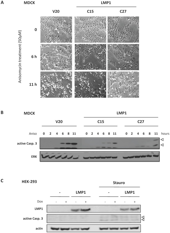
Figure 5. Cell lines stably expressing LMP1 were more resistant to stress-induced cell death.
(A, B) MDCK cell lines stably expressing LMP1 (clones C15 and C27) and a control clone (V20) were treated with anisomycin (Aniso), an inhibitor of translation known to induce apoptose. (A) Representative pictures of cells cultured in the presence or absence of anysomycin (light microscope, magnification x20). (B) Detection of caspase 3 activation by Western blot analysis. Whole cell extracts were prepared after different periods of anysomycin treatment. Proteins were then resolved by 10% SDS-PAGE before Western blot analysis using antibodies against cleaved (i.e active form) caspase 3. ERK2 detection was used as loading control. (C) HEK-293 cell line stably expressing LMP1 and a control clone were treated for 8 hours with staurosporin (Stauro, 3 µM). Whole cell extracts were prepared and proteins were resolved by 10% SDS-PAGE before Western blot analysis of cleaved caspase 3 (white arrow). Actin was used as loading control.

