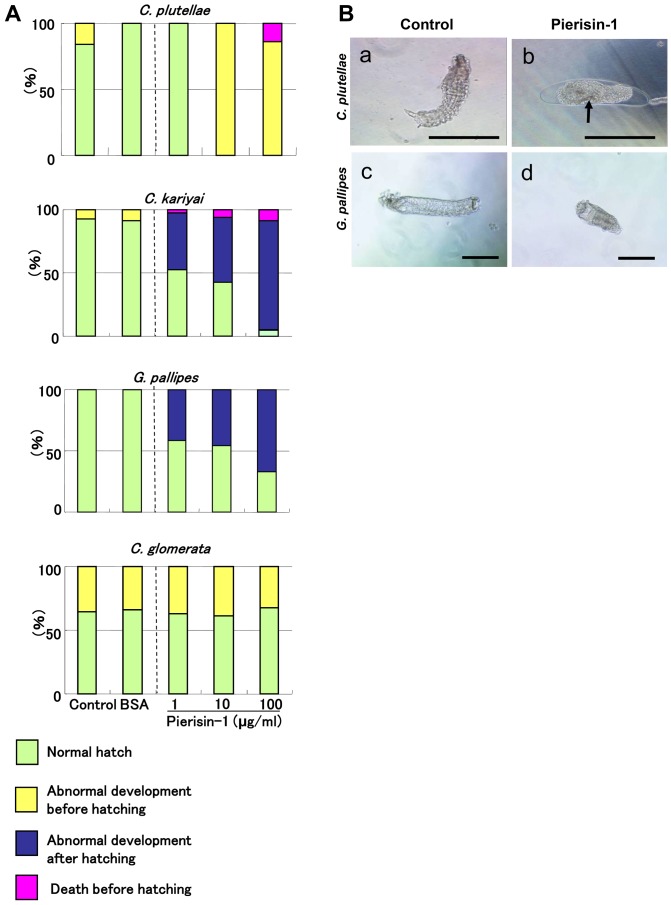
Figure 1. Effects of pierisin-1 on parasitoid eggs.
Segment formation completed parasitoid eggs were cultured for 2 days in media with or without pierisin-1. A: Rates of damaged eggs of parasitic wasps. Green column, normal hatch; yellow column, abnormal development before hatching; blue column, abnormal development after hatching; red column, death before hatching. Most C. kariyai and G. pallipes hatched in medium containing pierisin-1, but the larvae developed abnormally. Around 70% of C. glomerata hatched and developed normally. Some of them could not hatch but actively moved in the eggshell under these culture conditions. Therefore, we counted these wasps as “Abnormal development before hatching”. The numbers of eggs used for each treatment were 9 –13 for C. plutellae, 22 – 34 for C. kariyai, 10 – 26 for G. pallipes, 31 – 45 for C. glomerata. B: Phase contrast microscope images of cultured wasps. Embryo development of most C. plutellae was suppressed and cell death (arrow) was also observed with pierisin-1 treatment (a, b). G. pallipes larval length was shortened compared with the controls (c, d). Wasp eggs (n = 8 for each wasps) were treated with pierisin-1 at a final concentration of 10 µg/ml in the medium, and representative microscope images are shown. The anterior is on the left. Horizontal bars represent 200 µm.

