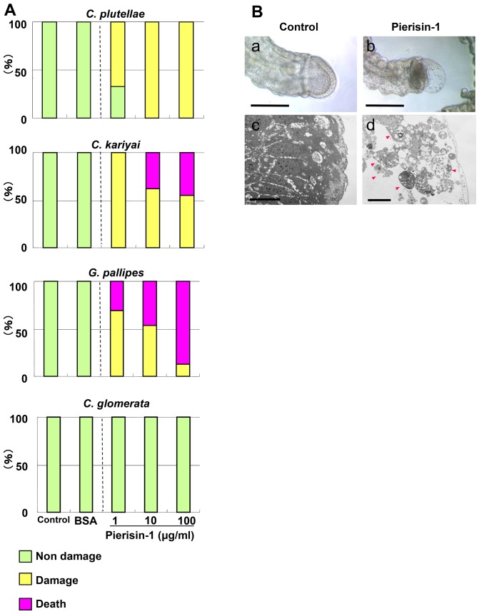
Figure 2. Effects of pierisin-1 on parasitoid larvae.
First instar larvae were cultured for 7 days in media with or without pierisin-1. A: Rates of damaged larvae of parasitic wasps. Green column, non-damage; yellow column, damaged; red column, death. C. glomerata larvae developed normally even in the presence of pierisin-1. Pierisin-1 treatment caused damage in larval bodies of non-habitual wasps. The numbers of larvae used for each treatment were 9 – 10 for C. plutellae, 11 – 16 for C. kariyai, 9 – 16 for G. pallipes, 11 – 25 for C. glomerata. B: Morphological analysis of cultured C. kariyai. a and b: Phase contrast microscope images of cultured C. kariyai larva with or without pierisin-1, respectively. c and d:. Transmission electron microscope (TEM) images of cross sections of their caudal vesicles. Apoptosis was observed in caudal vesicles on pierisin-1 treatment (arrowheads). Wasp larvae (n = 16) were treated with pierisin-1 at 10 µg/ml in the medium, and representative microscope images are shown. The anterior is on the left. Horizontal bars for phase contrast microscope represent 100 µm. Bars for TEM represent 1 µm.

