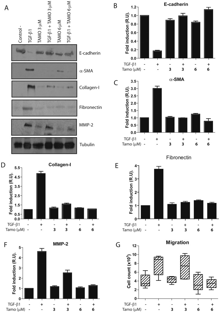
Figure 3. Tamoxifen blocks TGF-β1-induced MMT of MCs.
Omentum-derived MCs were treated or not with 1 ng/mL TGF-β1 for 24 or 48 hours, in the presence of different doses of Tamoxifen (0, 3 and 6 µM). (A) Western blot analyses show that Tamoxifen treatment prevents TGF-β1-induced E-cadherin down-regulation as well as α-SMA, collagen I, fibronectin and MMP-2 up-regulation. A representative experiment is shown. (B to F) The experiments were repeated at least five times and results are depicted as means ± SE. The expressions of E-cadherin (B) and MMP-2 (F) were analyzed at 24 hours, whereas the expressions of α-SMA (C), collagen I (D) and fibronectin (E), were analyzed at 48 hours of treatments. (G) Analysis of the migration capacity in transwell units demonstrates that Tamoxifen (6 µM) reduces the TGF-β1-indued migratory capacity of MCs to basal levels. The experiments, made in triplicates, were repeated at least four times. Box plots represent the median, minimum and maximum values, as well as the 25th and 75th percentiles.

