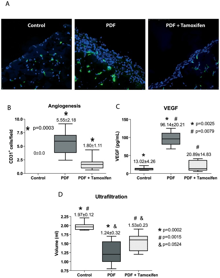
Figure 9. Treatment with Tamoxifen decreases PD-induced angiogenesis, inhibits VEGF production and improves peritoneal ultrafiltration.
Mice received a daily instillation of standard PD fluid with or without the oral administration of Tamoxifen (PDF; n = 14 and PDF + Tamoxifen; n = 17). A control group of mice that were instilled with saline was also included (Control; n = 9). (A) Standard PD fluid exposure increases peritoneal angiogenesis and Tamoxifen administration significantly reduces the number of vessels, as determined by CD31 staining (representative slides). Magnification ×200. (B) Box plots represent the CD31+ staining in the different experimental groups and show a decrease of angiogenesis in the Tamoxifen-treated animals. (C) Analysis of VEGF in the drained volumes shows a strong increase of this growth factor in PD fluid-instilled animals, and administration of Tamoxifen significantly reduces VEGF production. The ANOVA test resulted in a significance of p<0.0001. (D) A 30 minutes ultrafiltration test was performed on the last day of treatments. The volumes recovered from animals exposed to PD fluid are lower than those from mice instilled with saline solution and an increase of net ultrafiltration is obtained in mice exposed to PD fluid that were administrated Tamoxifen. A significance of p<0.0001 was obtained with the analysis of variance test. Box Plots graphics represent 25th and 75th percentiles, median, minimum and maximum values. Numbers above boxes depict means ± SE. Symbols represent the statistic differences between groups.

