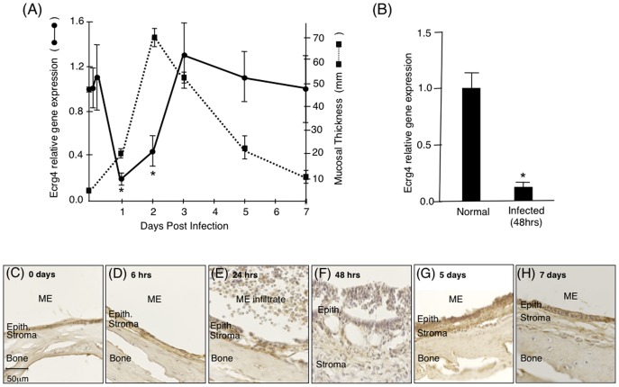
Figure 2. Characterization of changes in Ecrg4 gene expression in ME mucosal after NTHi infection.
Panel A: Mining a genechip microarray showed time-dependent decreased Ecrg4 expression levels after NTHi infection (solid line) of mouse ME. The decrease was compared to the thickness of the ME mucosa (dashed line). Mouse Ecrg4 expression is down-regulated within 24 hrs, while mucosal hyperplasia increases beginning 24 hrs after infection and peaking at 48 hrs. Ecrg4 expression also recovers just prior to return of the mucosa to normal thickness. Each gene expression data point represents gene arrays obtained from 2 independent sets of 20 C57BL/6J mice and expressed as fold change from the expression levels measured at time 0 hr (see [32], [34] for details). *P<0.05. Panel B: RT-PCR confirmed that Ecrg4 mRNA is expressed in normal rat ME mucosa and that it is down-regulated 48 hrs after NTHi infection. Bars represent the mean ± SEM (n = 4 MEs per time point). *Significantly different from normal (P<0.05). Panels C–H Immunohistochemistry of rat ear tissue harvested at 0 hrs, 6 hrs, 24 hrs, 48 hrs, 5 days, and 7 days after NTHi infection showed changes in Ecrg4 immunostaining in the ME mucosa. Epith. = epithelial cells.

