Figure 1.
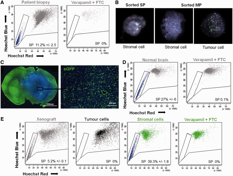
The side population in glioblastoma patient biopsies is non-neoplastic. (A) Side population (SP) discrimination assay performed on human glioblastoma: viable, single and nucleated cells (grey) of tumour biopsies (shown for T316) contained a visible side population (blue) identified as a well-defined characteristic ‘tail’ with a decreased Hoechst signal in both ‘Hoechst’ channels. Dye efflux was confirmed by double inhibition with 250 µM verapamil and 10 µM fumitremorgin C (FTC). Additional examples in Supplementary Fig. 2. (B) Fluorescence in situ hybridization analysis of side population and main population (MP) cells sorted from patient biopsies demonstrates the lack of tumour cells in the side population fraction (example shown for T330). Tumour cells showed typical glioblastoma aberrations [red probe = 10q23 (PTEN) deletion, green probe = EGFR amplification, blue probe = 7q trisomy]. (C) Glioblastoma spheroid-derived xenograft developed in the brain of enhanced GFP expressing NOD/SCID mice was recognized as a region with increased cellularity (DAPI = blue) and decreased enhanced GFP signal (green) (left). Higher magnification of the boxed area showing the presence of green host cells within the non-green human tumour tissue (right). (D) Normal mouse brain contained the side population phenotype (left), as confirmed by the inhibition control (250 µM verapamil + 10 µM fumitremorgin C) (right) (n = 8). (E) Side population phenotype was also detected in viable, single and nucleated cells of the patient-derived xenografts (T238) in enhanced GFP+ NOD/SCID mice (grey) (n = 3). Discrimination between host stromal (green) and tumour (black) compartment revealed side population uniquely in the stromal compartment of the xenograft (blue). Data are presented as a mean value ± SEM, n ≥ 4. For patient biopsies SEM represents technical replicates. See Supplementary Fig. 1 for detailed gating strategy and Supplementary Fig. 3 for additional examples of xenografts.

