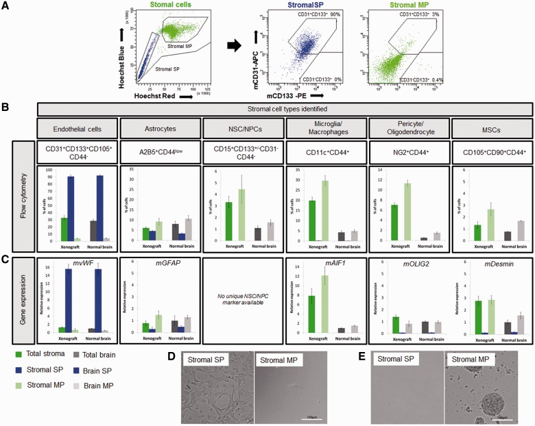
Figure 4.
Stromal cells with functional efflux properties within glioblastoma xenografts belong to endothelial and astrocytic populations. (A) Side population analysis (left) and CD133/CD31 phenotyping in the stromal compartment of the T16 xenograft (right). More than 90% of the stromal side population (blue) was CD31+CD133+, whereas CD31-CD133+ cells were devoid of a side population. Stromal main population (light green) contained 0.4% of CD31-CD133+ and only 3% of CD31+CD133+ events. (B) For identification of stromal cells, side population analysis was combined with multicolour phenotyping using antibodies against mouse epitopes and results were compared to normal mouse brain. Percentages of subpopulations of cells detected in the stromal compartment of the xenograft and normal brain were calculated for the whole single nucleated viable population (total stroma and total brain), side population (stromal side population and brain side population) and main population cells (stromal main population and brain main population) (n ≥ 4). See Supplementary Table 2 for cell membrane marker expression profile. Side population events were identified as CD31+CD133+CD105+CD44- endothelial cells (first panel) and A2B5+CD44low astrocytic cells (second panel). Other populations characterized did not contain side populations (panels 3–6). Note that the A2B5+ side population cells co-expressed the glial marker CD44 at a medium/low level and were therefore not included in the CD44+ populations in our calculations. (C) Expression of lineage-specific markers was analysed by quantitative real-time-PCR. Mouse-specific actin and GAPDH were used as reference genes. The data are presented as mean ± SEM (n = 3). Normal brain (total brain) was used as an internal calibration (value = 1). See Supplementary Table 2 for internal cell marker expression profile and Supplementary Fig. 4 for sorting controls. Unfortunately none of the neural stem/progenitor cell markers was specific enough for PCR analysis, since nestin, vimentin (Supplementary Fig. 7E), SOX2 and GFAP are also expressed by other cell types in the brain. Desmin was used as a marker of pericytes and mesenchymal stem cells. (D) When sorted stromal side population and stromal main population cells were cultured in endothelial cell medium, only cells from the side population fraction were able to survive under these conditions. Only at high cell concentrations (100 000/well) occasional endothelial cells present within the main population survived. (E) Under neural stem cell conditions neurospheres developed only from the main population fraction (10 000 cells/well). MP = main population; MSC = mesenchymal stem cells; NPC = neural precursor cell; NSC = neural stem cell; SP = side population.

