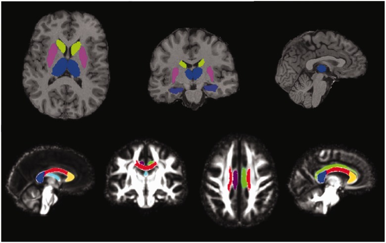
Figure 1.
Regions of interest. Grey matter regions of interest (top): segmentations of thalamus (blue), caudate (green), putamen (pink) and hippocampus (purple) overlaid on the T1 image of a single subject. White matter tracts of interest (bottom): segmentations of the fornix (pale blue), right cingulum (purple), left cingulum (green), genu (yellow), body (red) and splenium (dark blue) of the corpus callosum, overlaid on the study-specific template.

