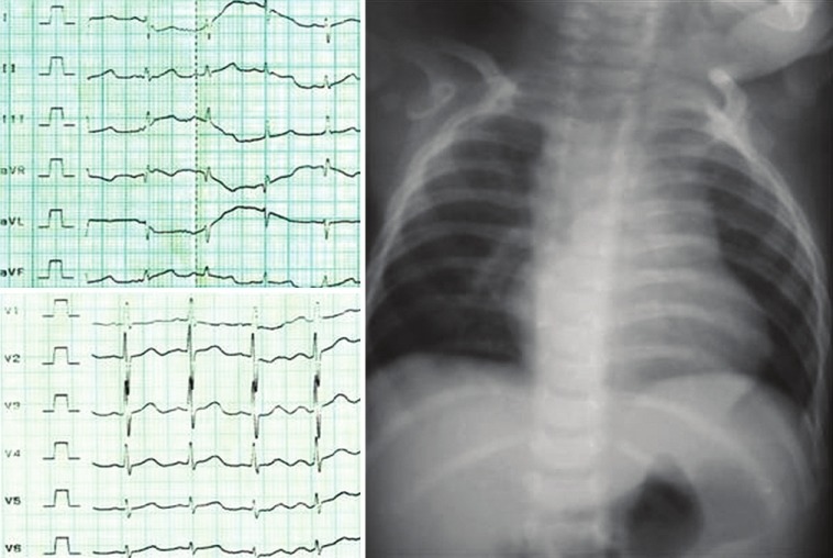
Figure 2.

Electrocardiogram (left side) showed normal sinus rhythm with right ventricular hypertrophy with right axis deviation. Chest roentogram (right side) showed levocardia, oligemia, cardiomegaly (cardiothoracic ratio of 60%) mainly of right ventricle, normal thoracic situs
