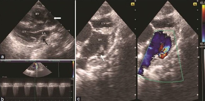
Figure 3.

Parasternal long axis view (a) demonstrating large subaortic ventricular septal defect with more than 50% over riding by aorta (thick white arrow) and dilated coronary sinus (black arrow). Subcostal view (c) demonstrating both great vessel committed to right ventricle with Doppler evidence of severe right ventricular outflow tract obstruction (thin white arrow) having peak gradient of 65 mm of mercury (b) (LV = Left ventricle;RV = Right ventricle;LA = Left atrium)
