Figure 1.

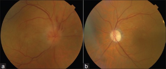
(a) Funduscopic examination revealed hyperemic and swollen optic disc with small hemorrhages in the right eye ( b ) Pale and atrophic optic disc in the left eye

(a) Funduscopic examination revealed hyperemic and swollen optic disc with small hemorrhages in the right eye ( b ) Pale and atrophic optic disc in the left eye