
Figure 3. NKp44+CD103+cells express T-bet, Eomes and Aiolos.
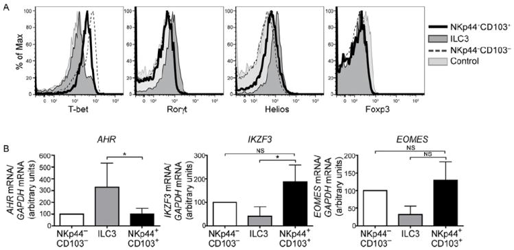
(A) Tonsil CD56+ cells were stained for intracellular T-bet, RORγt, Helios, and FoxP3. Dark gray profiles indicate ILC3, black lines represent NKp44+CD103+ ILC1, and dotted lines indicate NKp44−CD103− cells. Light gray profiles indicate staining with isotype-matched control antibody. Representative data from 3 individual tonsil samples are shown. (B) Tonsil CD56+ cells were sorted into the three subsets described in (A), and mRNA content for AHR, IKZF3 (encoding Aiolos) and EOMES was analyzed by qRT-PCR. Displayed are normalized data from 4 (AHR), 9 (IKZF3), and 5 (EOMES) donors. Data are represented as mean +/- SD.