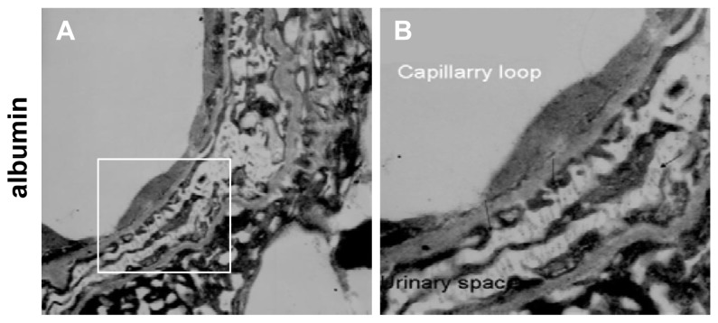
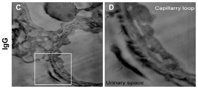
Figure 2.
Immune electron micrographs of albumin and IgG in the glomerular filtration membrane under acute hypertensive conditions. Under the acute hypertensive condition, the distributions of albumin (A,B) and IgG (C,D) were changed. Both albumin and IgG were clearly immunolocalized along the apical surface of the podocytes and Bowman’s spaces (arrows). In addition, podocyte fusion and reduced microvilli could be observed. (Magnification, ×8000 for A and C, ×12000 for B and D).


