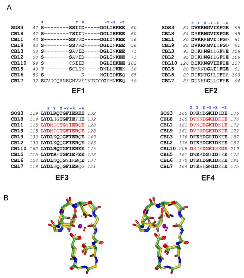
Figure 5.
The Ca2+ binding EF hands of CBLs. (A) Sequence alignment of the AtCBL EF hands. Canonical EF hands are shown in red and residues involved in Ca2+ binding are highlighted by X, Y, Z, -X, -Y, -Z according to a classical EF hand. (B) Stereo view of the superimposition of a classical EF hand Ca2+ binding loop (EF-2 of CnB, PDB code 1AUI) and SOS3 EF-4 (PDB code 1V1G).

