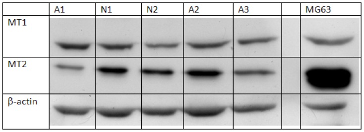
Figure 1.
Representative image of protein expression of melatonin receptors in osteoblasts. Cells isolated from normal controls were cultured until confluence. Cells were then collected and lysed for analysis of protein expression of melatonin receptor MT1 and MT2. Beta-actin was used as an internal control, and protein from the cell line MG63 was used for positive control. N = normal control; A = AIS subject.

