
Figure 4. Invisible InsP3-dependent sarcoplasmic reticulum (SR)-Ca2+ release unmasked by SR-Ca2+ leak-load relationship.
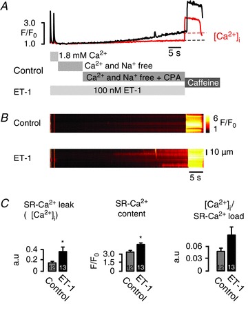
A, representative data showing averaged F/F0 of fluo-3 fluorescence changes (control: red; endothelin-1 (ET-1): black) and B, corresponding line scan images of fluo-3 fluorescence recorded in atrial myocytes. Cells were field stimulated for at least 45 s at 1 Hz before line scan images were recorded. After pre-pulse protocol, superfusion was rapidly switched from 1.8 mm[Ca2+]o to a nominally [Ca2+]o- and [Na+]o-free extracellular solution for 15 s, then 10 μm cyclopiazonic acid (CPA) was added for 20 s, before 10 mm caffeine was applied. The protocol was repeated in the presence of 100 nm ET-1 after pre-incubation with 100 nm ET-1 for at least 120 s. The increase in background fluorescence when CPA was applied represents SR-Ca2+ leak (Δ[Ca2+]i). C, averaged data, SR-Ca2+ leak (Δ[Ca2+]i), SR-Ca2+ content and SR-Ca2+ leak/SR-Ca2+ load relationship (Δ[Ca2+]i/SR-Ca2+ load) compared with control (no InsP3 pathway stimulation). In the presence of ET-1 there was a significant increase in the SR-Ca2+ leak (Δ[Ca2+]i) when CPA was added, unmasking a substantial contribution of the InsP3R opening to the total SR-Ca2+ leak. *P < 0.02–009 vs. control, N= 3; n= 12–13, mean ± SEM.