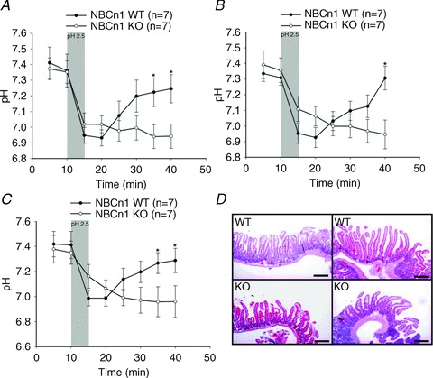
Figure 1. NBCn1 knock-out (KO) mice failed to recover after luminal acid-induced intracellular acidification.

A–C show time course experiments for enterocyte pHi measured at different distances from the villus tip (A, 100 μm; B, 200 μm; and C, 300 μm) in SNARF-1 AM-loaded villi of luminally perfused, exteriorized duodenum in anaesthetized NBCn1 WT and NBCn1 KO mice. Villous enterocyte pHi recovered quickly after the removal of acidic saline in WT duodenum, while almost no pHi recovery was observed in NBCn1 KO duodenocytes during the observation period. The shaded bars indicate the time period for the application of low pH. The numbers of mice are given in parentheses. *P≤ 0.05 between the groups. D, Haematoxylin and Eosin staining of the duodenum after the experiments, indicating that the short exposure of the mucosa to pH 2.5 did not result in discernible villous tip damage and that no histological difference was detected in the duodenum of NBCn1 WT and NBCn1 KO mice. Scale bars represent 500 μm.
