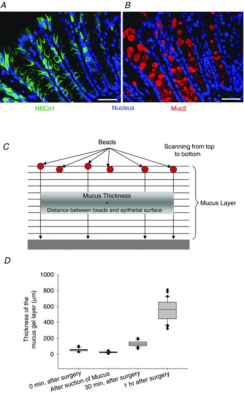
Figure 6. Colonic NBCn1 and mucin 2 (Muc2) staining pattern and method for mucus layer build-up.

Staining for NBCn1 (A) and Muc2 (B) in murine distal colon suggests expression in the same cell compartment. Scale bars represent 50 μm. C, schematic image of the fluorometric assessment of colonic mucus layer build-up on the exteriorized colonic mucosal surface in vivo. Fluorescent beads were allowed to settle onto the surface of the mucus layer, and laser scanning was performed in parallel planes down to the epithelial surface, which can be detected by reflection of light from the epithelial surface. The mean distance between each bead and the epithelium in the z-axis was taken as a measurement of the accumulated mucus layer. D, the box plots represent the thickness of the accumulated mucus at different time points during the study. After mounting the colonic mucosa in the perfusion chamber, a little loose mucus remains attached (leftmost bar). The next column indicates the thickness of the mucus when the loosely adherent layer was gently sucked off. This represents the mucus layer thickness of the firmly adherent layer. As time progressed, the thickness of the accumulated mucus increased.
