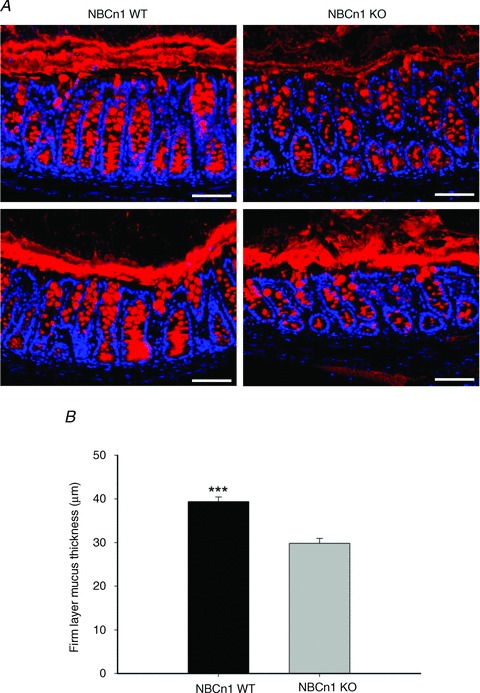
Figure 8. Assessment of the adherent mucus layer in Carnoy-fixed mid-distal colon.

A, Muc2 staining of the colonic mucus layer in NBCn1 KO and NBCn1 WT mice. Two examples of mucus layer staining for WT (two left panels) and for NBCn1-deficient mice (two right panels) are displayed, although the majority of images looked like the upper two images. Scale bars represent 75 μm. B, systematic assessment of the thickness of the mucus (see Methods) in sex- and age-matched NBCn1 KO and WT mice revealed a slight but significant reduction in mucus layer thickness in NBCn1 KO mid-distal colon compared with WT littermates. n= 3. ***P≤ 0.001
