
Figure 1. Representative laser scanning confocal immunofluorescence triple labelling of kidney cortex from aquaporin-2 (AQP2)-connecting tubule (CNT)-knockout (KO) mice (A, C and E) and control mice (B, D and F).
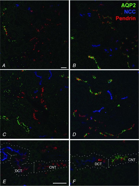
Each image is from a different animal. Late distal convoluted tubule (DCT), CNT and early cortical collecting duct (CCD) B-type intercalated cell marker pendrin is pseudo-labelled in red; DCT marker NCC is pseudo-labelled in blue; and AQP2 is pseudo-labelled in green. AQP2-CNT-KO mice have long segments of distal tubule solely immunolabelling for pendrin in contrast to the control mice having co-labelling of pendrin and AQP2 in similar segments, demonstrating deletion of AQP2 in the CNT. E, the transition from NCC-positive DCT to pendrin-positive CNT is indicated by the dotted line, and only control mice (F) show co-labelling with AQP2 in the pendrin-positive CNT. Scale bar: 200 μm.