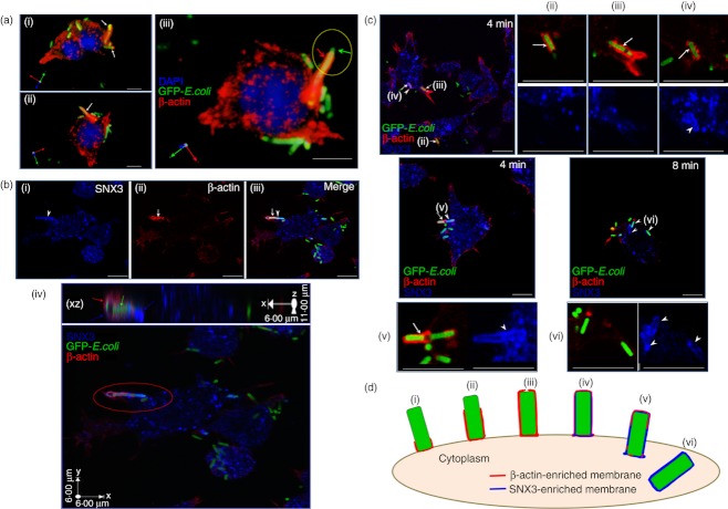
Figure 3.
Sorting nexin 3 (SNX3) recruits to β-actin-positive nascent phagosomes. (a) β-Actin is a marker for phagocytic cup. DC2.4 cells were pre-incubated with fixed GFP-Escherichia coli on ice for 1 h, after which they were washed to remove extracellular unbound GFP-E. coli. Cells were allowed to phagocytose the GFP-E. coli for 4 min at 37° before they were washed and fixed on ice. Cells were stained with antibody against β-actin (monoclonal), followed by Cy3-conjugated anti-mouse IgG. Stereo three-dimensional rendering of collected confocal images showing the recruitment of β-actin (red) to the GFP-E. coli-containing phagocytic cups (white and red arrows). (b) β-Actin and SNX3 co-localize at the nascent phagosomes. Following the same procedure for GFP-E. coli uptake, cells were co-stained with antibody against β-actin (monoclonal) and SNX3 (polyclonal), followed by Cy3-conjugated anti-mouse IgG and AlexaFluor®647-conjugated anti-rabbit IgG. (Panel i–iii) Confocal images of the co-localization of β-actin (red) and SNX3 (blue) at the nascent phagosomes. Arrowhead points to the complete SNX3 enrichment and arrow points to the partial β-actin enrichment at the nascent phagosome. (Panel iv) a stereo three-dimensional rendering and X–Z projection of the confocal images showing SNX3 localized to the whole length of the nascent phagosome (blue arrows) and β-actin localized to part of the nascent phagosome (red arrow). (c) Confocal images showing the complete enrichment of SNX3 to nascent phagosomes/early phagosomes, as well as the differential enrichment of β-actin to phagocytic cups/nascent phagosomes at different stages of phagocytosis. Arrowheads point to SNX3-positive nascent phagosomes/early phagosomes and arrows indicate β-actin-positive phagocytic cups/nascent phagosomes. Roman numerals (ii) – (vi) indicate the different stages of phagocytosis represented, corresponding to Fig. 3(d). Enlarged images are shown in corresponding panels (ii) – (vi). (d) A schematic showing the different stages during phagosome synthesis, namely phagocytic cup initiation (i), phagocytic cup extension [(ii) and (iii)], transitional closure (iv) and nascent phagosome separation from the plasma membrane [(v) and (vi)]. Scale bars: 10 μm.

