
Figure 1. The Thy1 Promoter Drives Transgenic Expression of ChR2-YFP in Subsets of Neurons in the Central Nervous System.
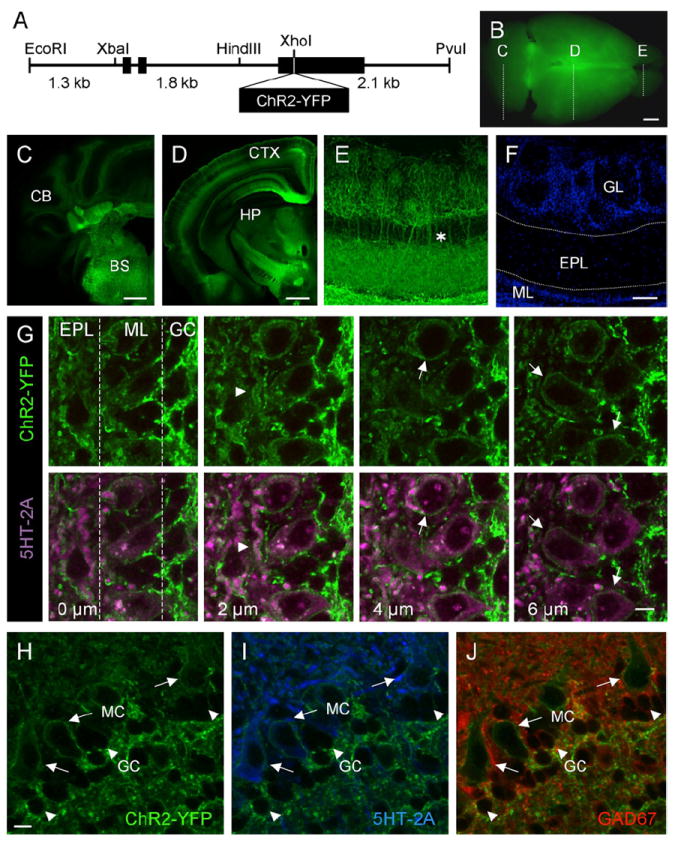
(A) Diagram of the Thy1-ChR2-YFP transgene construct.
(B) Whole-brain image of ChR2-YFP expression in Thy1 transgenic mouse brain, illustrating the coronal planes of section shown in (C)–(E).
(C) Coronal section through the caudal end of the Thy1-ChR2-YFP transgenic mouse brain. CB, cerebellum; BS, brainstem.
(D) Midcoronal section through the Thy1-ChR2-YFP transgenic mouse brain. CTX, cortex; HP, hippocampus.
(E) Immunoenhanced section using an anti-GFP antibody to amplify ChR2-YFP detection in the mitral cells of the olfactory bulb. The asterisk illustrates apical dendrites extending from mitral cell bodies, forming tufts in the glomerular layer.
(F) DAPI stain of the section shown in (E), highlighting the cell layers of the olfactory bulb. GL, glomerular layer; EPL, external plexiform layer; ML, mitral cell layer.
(G) Serial z sections taken at 2 μm intervals through the olfactory bulb of Thy1-ChR2-YFP mice showing the localization of ChR2-YFP at the plasma membrane of mitral cells. Top panels show ChR2-YFP expression in serial z planes of section. Bottom panels show corresponding planes of section with colabeling of mitral cell bodies and lateral dendrites with an antibody directed against the mitral cell-expressed 5-HT2A receptor (purple) and ChR2-YFP (green). The arrowhead points to ChR2-YFP expression on the membrane of a lateral dendrite. Arrows point to ChR2-YFP expression on the membrane of mitral cell bodies. EPL, External plexiform layer; ML, mitral cell layer; GC, granule cell layer.
(H–J) Within the olfactory bulb, ChR2-YFP is selectively expressed in mitral cells.
(H) High-magnification view of ChR2-YFP expression in the mitral cell layer.
(I) Colocalization of membrane-bound ChR2-YFP (green) and 5-HT2A receptor (blue), a marker of mitral cells.
(J) ChR2-YFP is not expressed in GAD67-positive granule cells (red). Note the presence of membrane-localized ChR2-YFP on the mitral cell (long arrows) with no ChR2-YFP present on the smaller GAD67-positive granule cell (arrowheads).
MC, mitral cell; GC, granule cell. Scale bars, 2.5 mm (B); 1 mm (C and D); 50 μm (E and F); 10 μm (G); 10 μm (H–J).