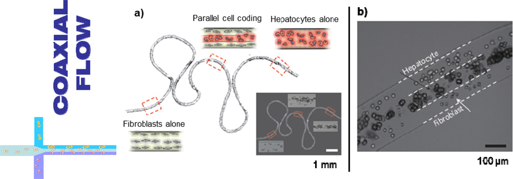
Figure 4.
(a) Schematic and micrograph (inset) of an alginate microfiber, spatially coded to include either fibroblasts, rat hepatocytes, or a mixture of the two cell types. In the first case the cells were coded into the fiber serially; in the latter case the coding was parallel; (b) Higher magnification micrograph of a fiber section containing the cell co-culture. Figures adapted and reprinted with permission from [136].

