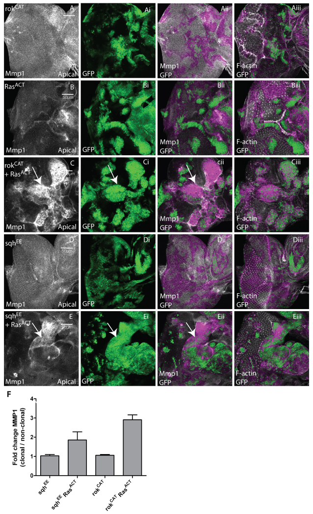
Fig. 8.
Activation of Rok and Myosin II lead to increased expression of the JNK target Mmp1 in EAD clones. Confocal planar apical sections through the epithelium of third instar larval EADs. Mutant clones are marked by the presence of GFP (green). EADs were stained for Mmp1 to reveal JNK activity (white) and with phalloidin-TRITC to detect F-actin (white). (A) rokCAT. (B) RasACT. (C) rokCAT + RasACT. (D) sqhEE. (E) sqhEE + RasACT. Third instar EADs from rokCAT mosaic larvae did not show upregulation of Mmp1 in the majority of clones, although expression was mildly induced in some clones (not shown). Expression of RasACT alone did not upregulate Mmp1 in the majority of clones. Mmp1 was strongly upregulated in most rokCAT + RasACT clones (arrows, C-Cii) compared with adjacent wild-type tissue. Expression of sqhEE did not lead to Mmp1 upregulation in the clones (D), whereas expression of sqhEE + RasACT resulted in induction of Mmp1 in many but not all clones (arrows, E-Eii). (F) Quantification of Mmp1 levels in sqhEE, sqhEE + RasACT, rokCAT or rokCAT + RasACT clones versus wild-type clones. The data was compared by a t-test and error bars represent s.e.m. The significance was P<0.0001 for rokCAT + RasACT compared with rokCAT and P<0.08 for sqhEE + RasACT compared with sqhEE.

