Fig. 9.
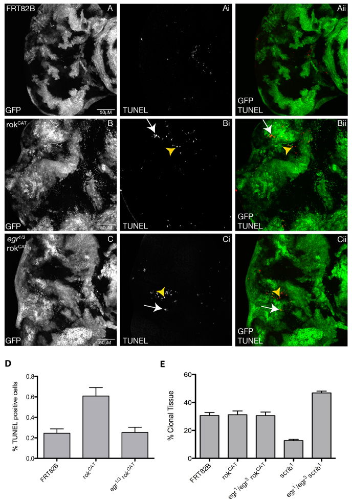
TNF (Eiger) is required for apoptosis in rokCAT clones. Confocal planar compiled sections through the epithelium of third instar larval EADs. Mutant clones are marked by the presence of GFP (white, and green in the merge) and TUNEL (white, and red in the merge). (A) FRT82B control. (B) rokCAT. (C) egr1/3rokCAT. Third instar EADs from rokCAT mosaic larvae show higher levels of apoptotic cells, as revealed by TUNEL, both cell autonomously (arrows, Bi,Bii) and non-cell autonomously (arrowheads, Bi,Bii). Removing egr reduced, but did not eliminate, apoptotic cells in the rokCAT EAD in both rokCAT (arrows, Ci,Cii) and wild-type clones (arrowheads, Ci,Cii). (D) Quantification of the number of TUNEL-positive cells in EADs of each genotype indicated. The data was compared by a t-test and error bars represent s.e.m. The significance was P<0.05 for rokCAT compared with the FRT control, and for rokCAT compared with egr1/3rokCAT. (E) Quantification of the percentage of mutant clonal tissue relative to the total EAD size for each genotype indicated. The data was compared by a t-test and error bars represent s.e.m. The significance was P<0.05 for scrib1 compared with egr1/3 scrib1, for scrib1 compared with FRT, and for egr1/3scrib1 compared with FRT.

