
Fig. 5.
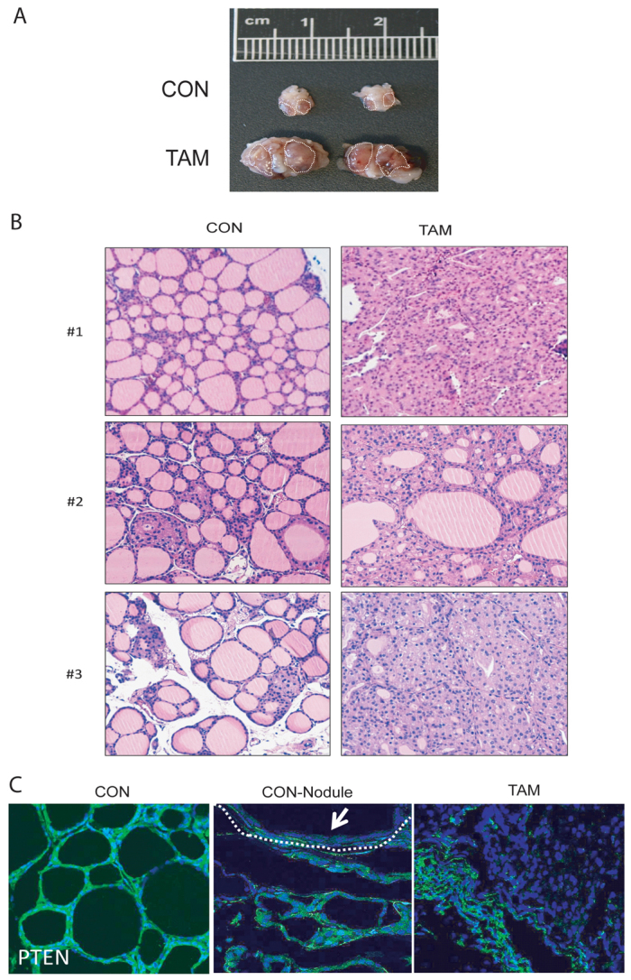
PTEN inactivation leads to the development of thyroid hyperplasia. (A) Comparison of representative thyroids from CAG-Cre-ERT+/−PTENfl/fl mice 6–8 weeks after being injected with corn oil (CON) or tamoxifen (TAM). (B) Representative images of hyperplasia in thyroids dissected from three corn-oil-injected (CON) and three tamoxifen-injected (TAM) mice. (C) Immunofluorescence staining showing PTEN loss in CAG-Cre-ERT+/−PTENfl/fl thyroids. Representative images showing PTEN immunofluorescence of mice injected with corn oil displaying no hyperplasic nodules (CON), mice injected with corn oil displaying hyperplasic nodules (CON-Nodule) or mice injected with tamoxifen (TAM). Arrow indicates PTEN-negative immunofluorescence in hyperplasic module.