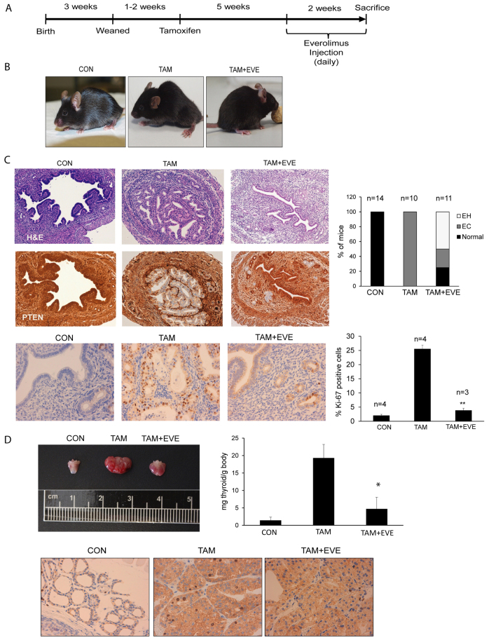
Fig. 7.
mTOR inhibition reduces endometrial lesions and thyroid hyperplasia. (A) Schematic representation of the protocol used for everolimus administration. Briefly, 5 weeks after tamoxifen injection, animals started to receive a single daily intraperitoneal dose of everolimus for 14 consecutive days and then they were sacrificed. (B) At 7 weeks after tamoxifen injection (TAM), mice exhibited lethargy, ruffled fur and hunched posture. Animals treated with everolimus (TAM+EVE) remained healthy and active, similar to controls (CON). (C) H&E staining and PTEN immunohistochemistry images corresponding to endometria from mice treated with corn oil (CON), tamoxifen (TAM) or tamoxifen plus everolimus (TAM+EVE). Everolimus-treated females showed reduced lesions and PTEN-negative healthy areas. Percentage of animals developing endometrial hyperplasia (EH) and endometrial in situ adenocarcinomas (EC) after injection of corn oil (CON), tamoxifen (TAM) or tamoxifen and everolimus (TAM+EVE) are shown in the top graph. Representative images (bottom left) and quantification (bottom right) of Ki-67 immunostaining performed on uterus dissected from mice injected with corn oil (CON), tamoxifen (TAM) or tamoxifen plus everolimus (TAM+EVE). (D) Representative images of thyroids dissected from mice injected with corn oil (CON), tamoxifen (TAM) or tamoxifen plus everolimus (TAM+EVE). Everolimus treatment also reduced thyromegaly in tamoxifen-injected mice. Everolimus caused a marked decrease of the index of proliferation, with levels similar to control animals, as assessed by Ki-67 immunostaining. *P≤0.05; **P≤0.001.

