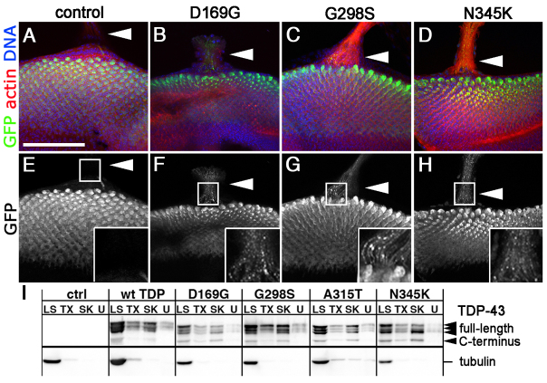
Fig. 2.
TDP-43 forms axonal aggregates in developing eyes. (A–H) Confocal images (single slices, 1 μm each or 2–3 slice projections) showing GFP NLS (A,E) or TDP-43 variant (B–D,F–H) localization when expressed in the developing eye neuroepithelium with GMR-Gal4. TDP-43 visualized via individual fluorescent YFP tags (indicated as GFP here). Filamentous actin labeled with phalloidin, DNA stained with Hoechst 33342 as indicated. Note TDP-43 puncta in axons (arrowheads in B–D,F–H and insets); compare with GFP NLS controls (arrowheads in A,E and inset). (I) Cellular fractionation of adult head tissue shows TDP-43 distribution complexes ranging from highly soluble (LS, low salt) to insoluble (U, urea). Triton X-100 (TX) and sarkosyl (SK) correspond to non-ionic and ionic detergent soluble fractions, respectively. TDP-43 was detected using anti-GFP antibodies. Tubulin was used as a loading control. Scale bar: 75 μm.

