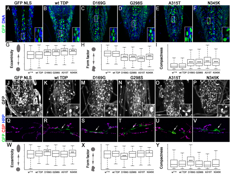
Fig. 3.
TDP-43 expression in motor neurons or glia leads to cytoplasmic aggregates and nuclear morphology defects. (A–F) TDP-43 expressed with D42 Gal4 was visualized using anti-GFP antibodies (B–F) and compared with D42 Gal4 >GFP NLS controls (A). Motor neurons within the ventral ganglia were labeled by GFP; DNA was stained with Hoechst 33342. (G–I) Mutant variants but not wild-type TDP-43 alter nuclear shape by increasing eccentricity (G), decreasing the form factor (H) and increasing compactness (I) compared with D42 Gal4 driver controls. (J–P) TDP-43 expressed in glia with repo Gal4 was visualized using anti-GFP antibodies (K–P) and compared with repo Gal4 >GFP NLS controls stained with Hoechst 33342 to visualize DNA (J). Note the presence of TDP-43 puncta in the cytoplasm (K–P). (Q–V) TDP-43 is expressed in puncta (arrows) within glial cells enveloping the neuromuscular junction synapse labeled by CSP and HRP. (W–Y) Glial expression causes relatively mild changes in nuclear shape, primarily in D169G and N345K compared with repo Gal4 driver controls. Eccentricity, form factor and compactness are shown as means ± s.e.m.; ***P<0.001. Scale bars: 20 μm.

