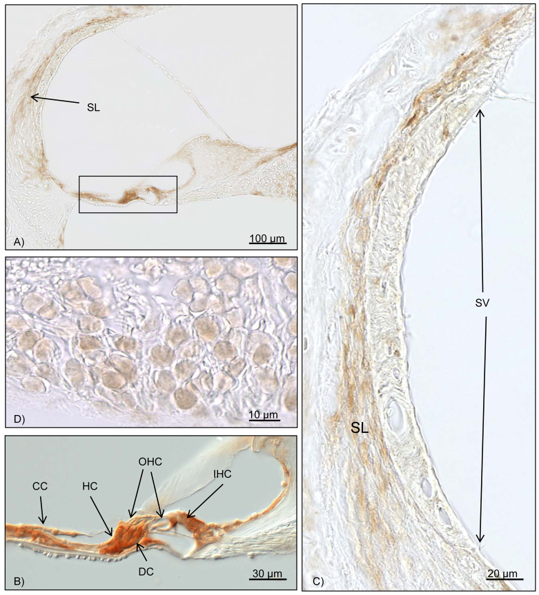
Fig. 4.
Immunohistochemical results for Bcl-2. Brown staining marks Bcl-2 activity. (A) Overview of the cochlea duct at the middle turn. The box marks the organ of Corti, with intensive Bcl-2 staining. Further reactivity can be seen in the spiral ligament. (B) Close-up of the organ of Corti. With the differential interference contrast mode, the inner and outer hair cells do not show any Bcl-2 activity. High Bcl-2 activation is seen for the Deiters’ cells, Hensen’s cells, Claudius’ cells and other supporting cells. (C) High magnification of the stria vascularis and the spiral ligament. Upregulation of Bcl-2 in the fibrocytes of the spiral ligament can be noted. No reaction is found in the stria vascularis. (D) Bcl-2 activation can be seen in the spiral ganglion cells. OHC, outer hair cell; IHC, inner hair cell; CC, Claudius’ cells; DC, Deiters’ cells; HC, Hensen’s cells; SL, spiral ligament; SV, stria vascularis.

