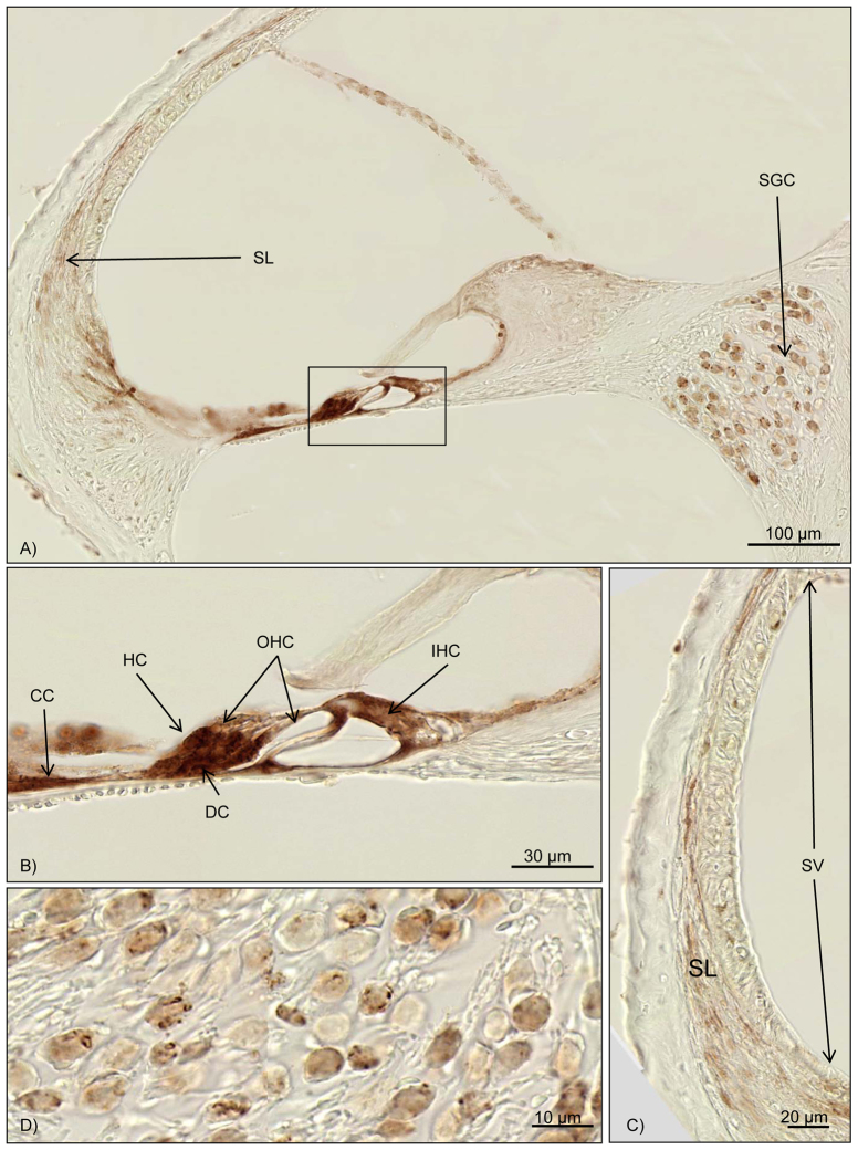
Fig. 5.
Immunohistochemical results for Bax. Brown staining marks Bax activity. (A) Overview of the cochlea duct at the middle turn. The box marks the organ of Corti, with intensive Bax staining. Further reactivity can be seen in the spiral ligament and the spiral ganglion cells. (B) Close-up of the organ of Corti. High Bax-2 activation is seen for Deiters’ cells, Hensen’s cells, Claudius’ cells and other supporting cells. (C) High magnification of the stria vascularis and the spiral ligament. Upregulation of Bax in the fibrocytes of the spiral ligament can be noted. No reaction is found in the stria vascularis. (D) Bax activation can be seen in the spiral ganglion cells. OHC, outer hair cell; IHC, inner hair cell; CC, Claudius’ cells; DC, Deiters’ cells; HC, Hensen’s cells; SGC, spiral ganglion cells; SL, spiral ligament; SV, stria vascularis.

