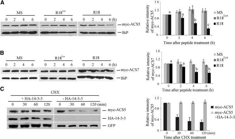
Figure 2.
14-3-3 Positively Regulates ACS Protein Stability.
(A) and (B) R18 peptide results in a decrease in the steady state level of myc-ACS5 (A) or mycACS7 (B) proteins. Seedlings expressing myc-ACS5 or myc-ACS7 were treated with 10 µg/mL R18, R18Lys, or an MS medium control for the indicated times. Total protein extracts from these seedlings were then analyzed by immunoblotting using an anti-myc antibody or an anti-BiP antibody as a loading control. On the right is shown a quantification of the protein levels from three independent biological replicates per treatment using ImageJ software. The myc-ACS5 or myc-ACS7 bands were normalized to the BiP control, and these values were than normalize to the time zero, which was set to 1. Different letters indicate significant differences at P < 0.01 (analysis of variance, Tukey HSD post-hoc test); error bars = se.
(C) The half-life of ACS5 protein is increased by 14-3-3. Mesophyll protoplasts were transformed with a plasmid expressing myc-ACS5 with or without a plasmid expressing 14-3-3ω. At time 0, cycloheximide (CHX) was added (250 µM Cf) and total proteins extracted at the indicated times and then analyzed by immunoblotting using the indicated antibodies. The myc-ACS5 signal was normalized to the GFP signal, and these values expressed relative to the time 0 value, which was set to 1. A quantification of the protein levels from three independent biological replicates is shown on the right. Values are presented as the mean ± se.

