Figure 2.
Mutations at PAT10 Caused Pollen Developmental Defects.
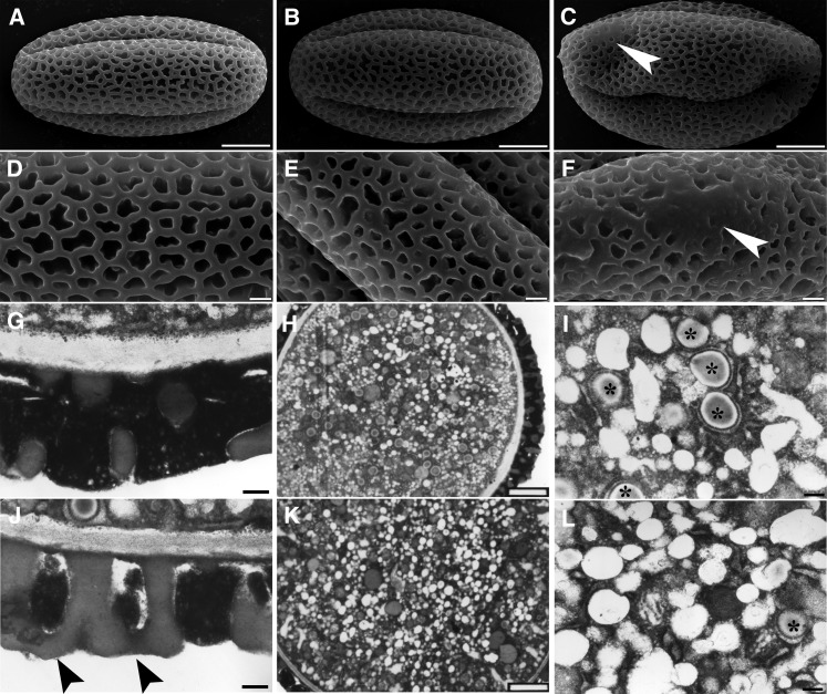
(A) to (C) Scanning electron micrographs (SEM) of mature pollen from the wild type (A), heterozygous pat10 mutants (B), and homozygous pat10 mutants (C). Arrowhead indicates defective pollen coat.
(D) to (F) Close-ups of SEM images shown in (A) to (C), respectively. Arrowhead indicates defective pollen coat.
(G) and (J) Transmission electron micrograph (TEM) of mature pollen from the wild type (G) and pat10 (J) showing pollen coat structure. Arrowheads indicate defective pollen coats.
(H) and (K) TEM section of mature pollen from the wild type (H) and pat10 (K).
(I) and (L) Close-ups of TEM shown in (H) and (K), respectively. Asterisks indicate lipid bodies.
Bars = 5 µm in (A) to (C), 1 µm in (D) to (F), 2 µm in (H) and (K), and 200 nm in (G), (I), (J), and (L).

