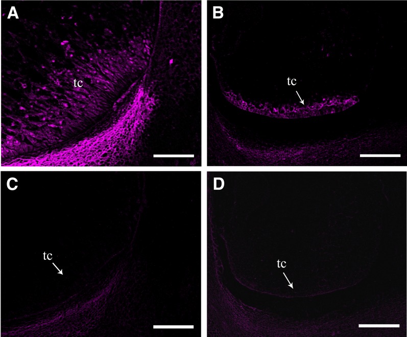
Figure 2.
The Basal Transfer Cell Development in the emp5-1 Kernels Is Arrested.
Confocal fluorescent microscopy visualization of BETL-2 by immunofluorescence in 13-DAP wild-type ([A] and [C]) and emp5-1 mutant ([B] and [D]) kernels. To visualize the basal endosperm transfer layer, specific antibody BETL-2 was used ([A] and [B]), and no BETL-2 antibody PBS buffer was used as control ([C] and [D]). tc, transfer cell. Bars = 200 µm.

