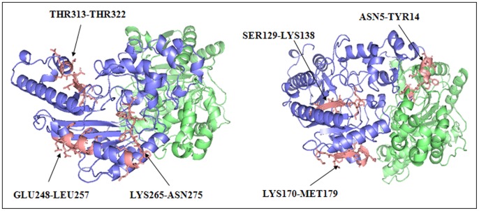
Figure 4. Positions of the peptides screened out by the peptide array membrane method on the HCK 3D structure.
The six experimental positive peptides were colored in salmon and represented by using the stick model. As shown in the picture, ASN5-TYR14 is at the position of dimerization interface of the two monomers; SER129-LYS138 is completely embedded inside the structure; GLU248-LEU257 is in a dent of the surface; LYS265-ASN274 which contains V8 endoproteinase fragment was also in a dent of the structure; LYS170-MET179 is located at the bottom of the structure; THR313-THR322 is located in the largest groove of the structure.

