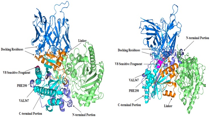
Figure 5. The scFv-A4-HCK complexes.
Model I is shown at left. Model II is shown at right.ScFv-A4 is colored in marine. HCK is shown as dimmer. One HCK monomer is colored in lime. The N-terminal of the other HCK monomer is colored in slate and C-terminal in cyan, the linker is colored in orange. The V8 endoproteinase sensitive fragment is colored in magentas. The binding residues are represented by using the stick model. The PHE250, VAL347 are represented using the sphere model.

