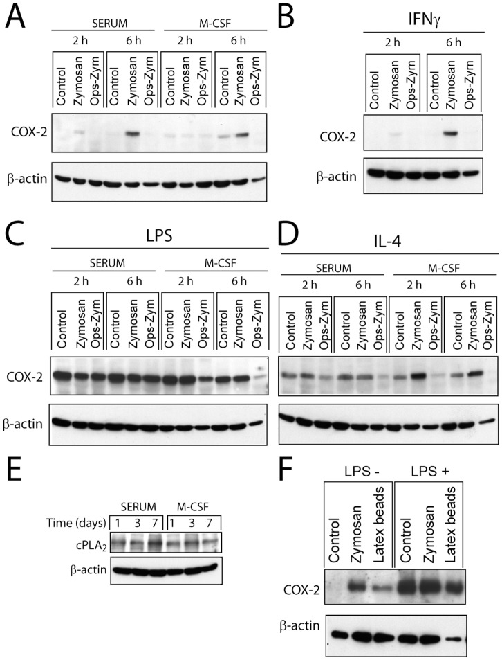
Figure 4. Expression of COX-2 protein.
(A–F) Macrophages were differentiated in the presence and absence of M-CSF and then primed with (B) 100 U/ml IFNγ, (C and F) 10 ng/ml LPS, and (D) 500 U/ml IL-4 for 3 hours. At the times indicated after stimulation, cell lysates were collected for the immunodetection of COX-2 and cPLA2 proteins. These are representative of experiments conducted at least in duplicate. (E) The expression of cPLA2 was assayed in macrophages differentiated in the presence of serum and M-CSF. (F) Macrophages were stimulated with latex beads at a concentration of 60 particles per cell.

