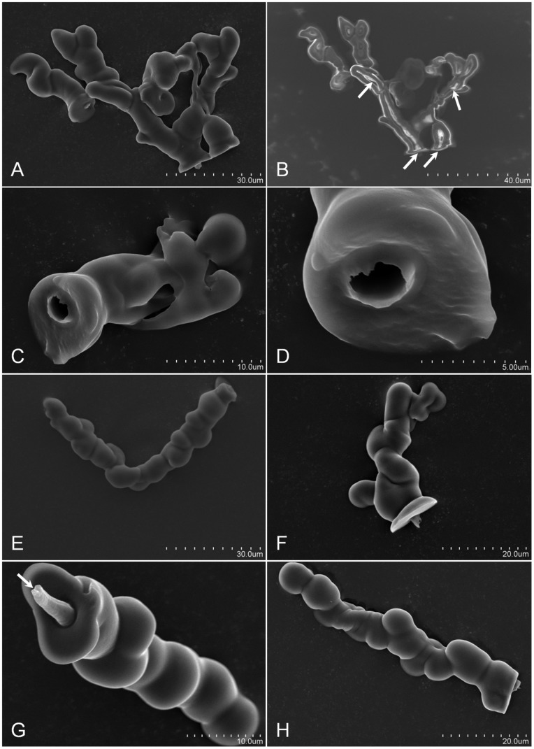
Figure 1. Scanning electron micrographs of D.citri in a¯ere stylet sheaths.
Panel A, is an image of two sheaths, the first (left sheath) is single-canal and the second (right) is a multi-branched sheath. Panel B is a non-gold sputtered image of Panel A with the white arrows indicating internal hollow canal tracks for the D. citri stylets. Panels C and D indicate the hollow canal of the D. citri stylets track with Panel D being an enlargement Panel C stylets canal opening. The opening is ∼3 µm in diameter. Panels E and F, indicate angular stylets probing (in āere) with panel E having a ∼93° bend within a single-canal sheath. Panels G and H are typical of linear sheaths formed in āere with panel G (white arrow indicating a closure of the stylet canal) suggesting secretion of sheath material as the stylet was retracted from the sheath.

