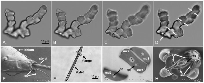
Figure 2. Micrograph images of D.citri in a¯ere formed stylet sheath and flange, and D. citri labrum and stylet bundle.
Panels A – C are individual confocal slices, with Panel D being a merged composite image of Panels A – C confocal slice images. Panel D, white arrows, indicate the visible internal stylet canal traversing the central core of the stylet sheath extending from the base to the terminal end of the sheath. Panel E is a SEM micrograph of a D. citri labium, stylets, and labial tip sensilla (ss). Panel F is a single slice confocal DIC micrograph of a D. citri third instar nymph exuvial stylet bundle with a detached flange circumambient the stylets. Panel G is a TEM micrograph cross-section of an adult D. citri stylet bundle indicating interlocking maxillary (mx) with mandibular (md) stylets, salivary (sc), food (fc), and dendrite (dn) canals. Panel H is a SEM micrograph of an adult D. citri flange formed in a mock feeding chamber. White arrows designate the location of indentations in the flange are sensilla cavities (ssc) with the central protrusion (black arrow) indicating a mound of retraction secreted sheath (rss) material formed upon withdrawal of the stylets from the sheath.

