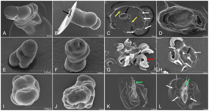
Figure 4. H.vitripennis, B. tabaci, and A. nerii in āere formed sheaths and flanges.
Panels A – D, E – H, and I – L are sheath and flanges from H. vitripennis, B. tabaci, and A. nerii, respectively. Panels A, B, E, F, I, and J are sheath and Panels C, D, G, H, K, and L are flange SEMs, respectively. For each, the sheaths are short and lack the branching as seen previously for D. citri, P. pyriformis, and F. virgata (for comparison see Figure 1 - Panels A and B, Figure 2 - Panel D composite, and Figure 4 - Panels B and E). Flanges for both the H. vitripennis (Panel C) and B. tabaci (Panel H) indicate an open access point into the central canal (designated by the black arrows) for the stylets. Panel D is an increased magnification SEM of the H. vitripennis flange opening for the stylets indicating the hollow canal for the stylets. Panels K and L indicate that the A. nerii flange are sealed closed by retraction secreted sheath material (green arrows). White arrows indicate sensilla cavities in the flange surface (Panels C, H and L), yellow arrows indicate the labial groove imprint on the H. vitripennis flange surface (Panel C), the red arrow indicating the coalescence of secreted whitefly ‘waxy’ cuticular lipid hydrocarbons shed and covering the flange (Panel G), and the black arrows (Panels B and H) indicate the open cavity of the stylet canal for both the H. vitripennis and B. tabaci flanges.

