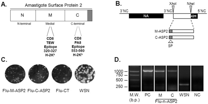
Figure 1. Construction and characterization of recombinant influenza viruses.
Schematic representation of primary sequence of Amastigote Surface Protein 2 and its corresponding moieties, highlighting the mapped CD8 T cells epitopes (A). Schematic representation of the neuraminidase dicistronic segment. NA38 segment contains an A/WSN/33 (WSN) derived recombinant neuraminidase (NA) segment followed by a duplicated 3′non coding (NC) sequence, XhoI and NheI cloning sites, a duplication of the last 42 nucleotides of NA (dark box) and the original 5′NC sequence (28 nucleotides). The foreign sequences (open boxes) were cloned between XhoI and NheI cloning sites (B).The plaque phenotype of the wild type WSN and recombinant influenza viruses were assessed by standard agarose plaque assay in MDCK cells after 3 days of incubation at 35°C and 5% CO2 (C).The NA segments of recombinant influenza viruses were analyzed by RT-PCR, using a set of primers that allows the amplification of the region containing the inserted foreign sequence. Corresponding plasmids DNAs were amplified in parallel as positive control. The amplified products were analyzes on a 1% agarose and visualized by ethidium bromide staining. The values depicted at the weight marker lane ar (D). W.M: weight marker; M: medial moiety of ASP2, C: carboxi-terminal moiety of ASP2; b.p.: base pairs.

