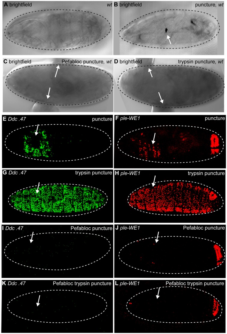
Figure 2. Serine proteases are required and sufficient for Ddc.47 and ple-WE1 activation.
Bright field images of wild-type stage 15–17 embryos. Ddc.47 and ple-WE1 are fluorescent reporters that include wound-induced DNA enhancers from the Ddc and ple loci, respectively. (A, C, D) A melanized wound site is not observed in unwounded, Pefabloc wounded, or trypsin wounded embryos. (B) Melanization at the wound site occurs after puncture-only wounding of wild-type embryos. Confocal images of Ddc.47 and ple-WE1 embryos 6 hours post wounding. (E, F) Control puncture, water puncture, and HCl (trypsin buffer) puncture wounded Ddc.47 and ple-WE1 embryos all exhibit localized reporter activation at epidermal wound sites. (G, H) Trypsin puncture wounded Ddc.47 and ple-WE1 embryos exhibit global reporter activation. (I, J) Pefabloc puncture wounded Ddc.47 and ple-WE1 embryos do not activate reporter at the wound site. (K, L) Pefabloc trypsin puncture wounded Ddc.47 and ple-WE1 embryos do not activate any epidermal wound reporter expression. The anal pad expression provided by the enhancer in the ple-WE1 wound reporter controls for developmental stage. Arrows mark the wound site. Dashed lines in the data panels mark the outlines of embryos.

