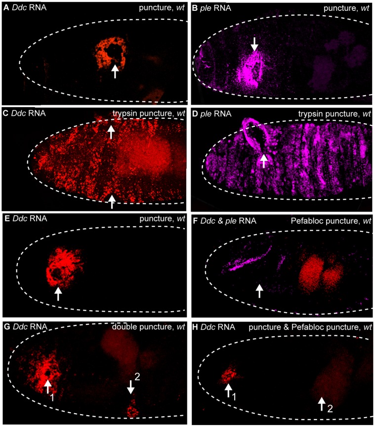
Figure 5. Serine proteases are sufficient and required for epidermal wound response gene transcription.
Confocal images of wild-type embryos after in situ hybridization with fluorescently labeled RNA probes made to detect transcripts from ple (magenta) and Ddc (red). (A, B) 30 minutes after HCl (trypsin buffer) puncture wounding, Ddc and ple transcripts accumulate in the epidermis around the wound site. (C, D) 30 minutes after puncture-trypsin wounding, ple and Ddc transcript accumulation can be observed throughout a large region of the epidermis. (E) One hour after water puncture wounding, wild-type embryos activate Ddc transcripts in the epidermis surrounding the wound site. (F) One hour after Pefabloc puncture wounding, no Ddc transcripts are activated in the epidermis surrounding the wound site in wild-type embryos, but normal developmental expression of ple in the cells that secrete the head skeleton is observed (magenta). Gut autofluorescence is seen in red. (G) In wild-type, double water puncture wounded embryos, 60 minutes after the first water puncture wound, a moderately wide zone of Ddc transcripts in the epidermis around wound sites is observed, while 30 minutes after the second wound, a narrow zone of Ddc transcript accumulation is observed around the wound site. (H) In wild-type double puncture wounded embryos with Pefabloc injected at the second site, 60 minutes after the first puncture wound, a narrow zone of accumulation of Ddc transcripts in the localized epidermis is observed around the first wound site, while 30 minutes after the second puncture wound with Pefabloc, no Ddc transcript accumulation is observed at the wound site. Arrows mark the wound sites. “1″ and “2″ indicate first and second wounds, respectively. Dashed lines in the data panels mark the outlines of embryos.

