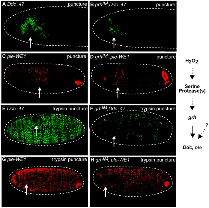
Figure 7. Serine protease-mediated wound reporter activation is upstream of grainyhead function.
Confocal images of control Ddc.47 and grhIM mutants; Ddc.47 or grhIM; ple -WE1 activation about six hours after puncture and trypsin puncture wounding. Ddc.47 and ple-WE1 are fluorescent reporters that include wound-induced DNA enhancers from the Ddc and ple loci, respectively. (A, B) Ddc.47 embryos puncture wounded with carrier solution activate localized reporter at the wound site, but dramatically reduced localized reporter activation is observed in grh mutants after the same treatment. (C, D) Ple-WE1 embryos puncture wounded with carrier solution activate reporter around the wound site, while grh mutants exhibit only slightly reduced reporter activation at the wound site. The developmental anal pad expression from the ple-WE1 reporter construct is observed in each treatment. (E, F) Puncture-trypsin wounded Ddc.47 embryos activate reporter globally, while grh mutants exhibit dramatically reduced and scattered wound reporter activation after trypsin treatment. (G, H) Trypsin-treated ple-WE1 embryos activate reporter globally, while grh mutants activate lower, patchier, but still easily detectable global reporter activation after trypsin treatment. Developmental ple -WE1 anal pad expression is observed in every treatment. The pathway is shown on the right side of the figure. Arrows mark the wound site. Dashed lines in the data panels mark the outlines of embryos.

