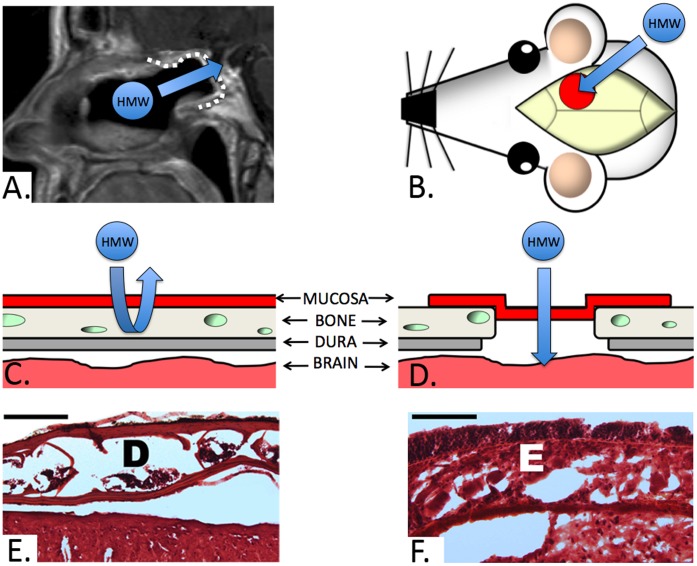
Figure 1. Murine graft model.
A) Sagittal MRI of a patient following endoscopic reconstruction of a skull base defect using a nasal mucosa graft(dotted white line, arrow denotes the proposed transmucosal pathway for HMW agents from the nose into the CNS through the graft). B) Illlustration of the murine graft model with the position of the graft(red circle) relative to the skull. The arrow denotes the equivalent transmucosal pathway to that seen on the MRI(Fig. 1A) utilized in our study. C and D) Cross sectional illustration of the skull base layers prior to and following craniotomy with dural removal and mucosal graft inset, respectively. Note that the dural layer(dura and arachnoid) contains the blood-cerebrospinal fluid barrier which restricts the transport of HMW molecules. E) Hematoxylin and eosin(H&E) section of the intact murine parietal bone with typical appearance of the inner and outer cortical tables with their associated diploic space(D) prior to engrafting(bar = 200 µm). F) H&E section of the mucosal graft implant in direct continuity with underlying brain parenchyma. Note the intact epithelial layer(E) consisting of pseudostratified columnar epithelium.

