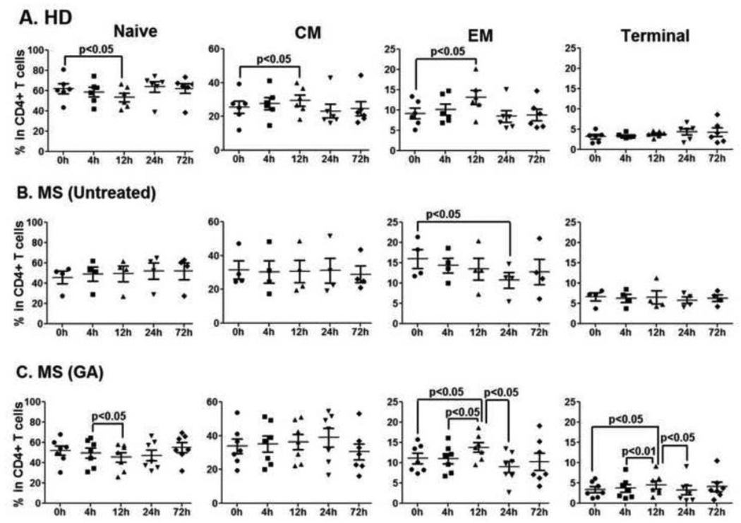
Figure 3. No GA-induced changes in CD4+ T cell compartment during first 72h of therapy.
Fluorescent conjugated anti-human CD27 and CD45RO were used for flow cytometry analysis to further differentiate CD4+ T cells into naïve (CD27+CD45RO−), central memory (CM, CD27+CD45RO+), effector memory (EM, CD27− CD45RO+) and terminal (Ter, CD27−CD45RO−) populations (Suppl. Fig. 1). Panels A, B and C represent percentages of naïve, CM, EM and terminal CD4+ T cells in HD, untreated, and GA-treated MS patients, respectively. Except for the terminal CD4+ T cells, similar diurnal fluctuations in CD4+ T cell subpopulations were present in all the study groups.

