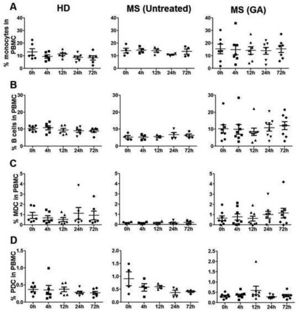
Figure 9. Percentages of monocytes, B cells, myeloid (MDC) and plasmacytoid dendritic cells (PDC) remain stable during first 72h of GA therapy.
Flow cytometry analysis using CD14, CD19, BDCA1 (MDC), and BDCA4 (PDC) anti-human antibodies was done in the peripheral blood of all the study participants throughout the observation time points. Data point in panels A, B, C & D represents % monocytes, B cells, MDCs and PDCs, respectively, in HD (n=6), untreated (n=4) and GA treated (n=8) MS patients.

