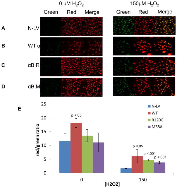
Fig. 4. Over-expression of wt αB-crystallin, αB-crystallin R120G and αB-crystallin M68A mutant proteins protects RPE mitochondria against oxidative stress damage.
Confocal microscopic images of RPE cells overexpressing control N-LV (A), wt αB-crystallin (B), αB-crystallin R120G (C), and αB-crystallin M68A (D) treated with 0 μM or 150 μM H2O2 for 24 h in serum free media. Cells were stained with JC-1 for 20 min to detect changes in mitochondrial membrane potential (MMP) as indicated by red mitochondrial staining (increased potential) or green cytosolic staining (decreased potential). This is reflected in the merged image as increased yellow fluorescence indicating decreased MMP. (B) Representative bar graph indicating the red/green ratio calculated from the mean intensities of three different fields for each experiment. Error bars represent standard deviation and p-values were calculated using the students t-test (n=3). Differences between intensity of the control N-LV and the αB-crystallin overexpressing cell lines were determined. p < 0.05 was considered statistically significant.

