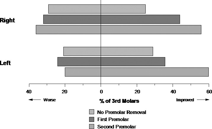
Figure 2.

Changes in mandibular third molar angulation from beginning to end of orthodontic treatment: no premolars removed (n = 24), first premolars removed (n = 25), and second premolars removed (n = 25). Improved indicates third molars were more vertical at the end of treatment as compared to the beginning of treatment.
